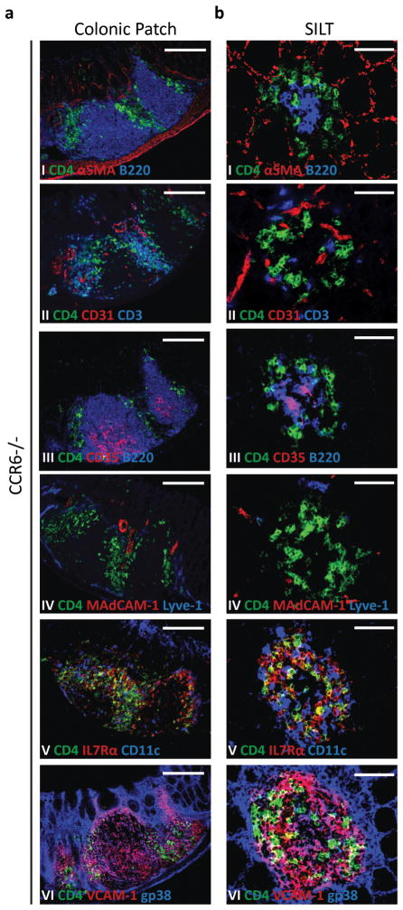
Figure 5. Colonic Patch and colonic SILT development is independent of CCR6.
Histological characterization of the colonic patches (a) and SILTs (b) present in the colon of 14 days-old CCR6−/− mice. Colon’s serial sections were stained for: (I) CD4 (green), αSMA (red), and B220 (blue); (II) CD4 (green), CD31 (red), and CD3 (blue); (III) CD4 (green), CD35 (red), and B220 (blue); (IV) CD4 (green), MadCAM-1 (red), and Lyve-1 (blue); (V) CD4 (green), IL7Rα (red), and CD11c (blue); and (VI) CD4 (green), VCAM-1 (red), and gp38 (blue). Scale bars (c) 100μm and (d) 50μm. At least 3 colons were entirely sectioned and the detected lymphoid tissues further analyzed.

