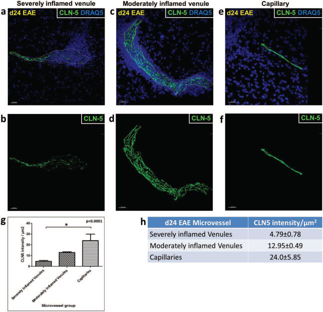
Fig. 3. CLN-5 density in spinal cord microvessels during EAE.
Isosurface rendering of the CLN-5 channel was performed in confocal z-stacks of spinal cord microvessels at d24 EAE. Top row, shows CLN-5 (Green) and nuclei/DRAQ5 (Blue) to highlight the close association of altered CLN-5 with dense perivascular cellularity. Bottom row, shows staining of only CLN-5 to emphasize significant TJ protein disruption. Inflamed venules demonstrated heterogeneity in CLN-5 loss: (a,b) severely inflamed venules displayed diffuse and extensive disruption of CLN-5; (c,d) moderately inflamed venules showed small punctate regions of CLN-5 loss; and (e,f) capillaries adjacent to severely inflamed venules appeared refractory to CLN-5 loss. 3D quantification of intercellular CLN-5 staining showed a significant reduction in intensity of CLN-5 staining per unit area of the endothelium in the severely inflamed venules compared to the capillaries (g,h). The boundaries of inflamed venules are marked with dashed white lines. A total of 6 microvessels were analyzed in each group sampled from 3 mice. *p < 0.0001. Scale bar = 20µm.

