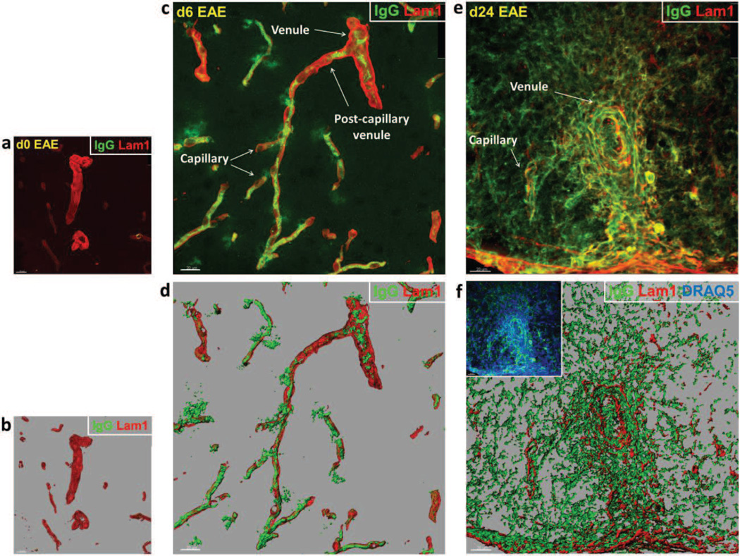
Fig. 6. Endogenous serum IgG leakage from spinal cord microvessels during EAE.
(a,c,e) Shows volume rendered images of confocal z-stacks from microvascular segments obtained from naïve mice, and mice at d6 and d24 EAE. (b,d,f) Shows the corresponding isosurface rendered images for purpose of enhanced spatial perspective. Staining of IgG (Green) and basement membrane/LAM 1 (Red) highlights vascular permeability around venules and capillaries. (a,b) Microvessels from naïve mice reveal no visible IgG immunostaining associated with venules or capillaries. (c,d) Microvessels at d6 EAE – prior to evidence of clinical disease – display focal IgG immunoreactivity around both venules and capillaries. (e,f) Microvessels at d24 EAE show pronounced and diffuse IgG immunoreactivity – reflecting endogenous serum protein extravasation – which obscured boundaries between the microvessel segments. Increased perivascular cellularity, indicative of leukocyte infiltration (inset), is highlighted by DRAQ5 staining (Blue). Scale bar = 20µm.

