
Figure 4. PAX6 is an early marker of pineal development and is only detectable at low levels in the adult.
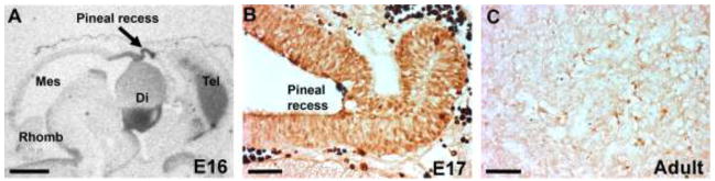
A) In situ hybridization for detection of Pax6 mRNA in a sagittal section of the rat brain at E16. Note the strong signal in the epithelium around the pineal recess (arrow). Di, diencephalon; Mes, mesencephalon; Rhomb, rhombencephalon; Tel, telencephalon. Scale bar, 1 mm. B) Immunohistochemical detection of PAX6 protein in the developing rat pineal gland at E17. Scale bar, 50 μm. The fetal animal was not perfused; therefore, the very dark cells are erythrocytes with a high level of endogenous peroxidase. C) Immunohistochemical detection of PAX6 protein in the pineal gland of a perfusion fixed adult rat. Scale bar, 50 μm. For methodological details, see [76].