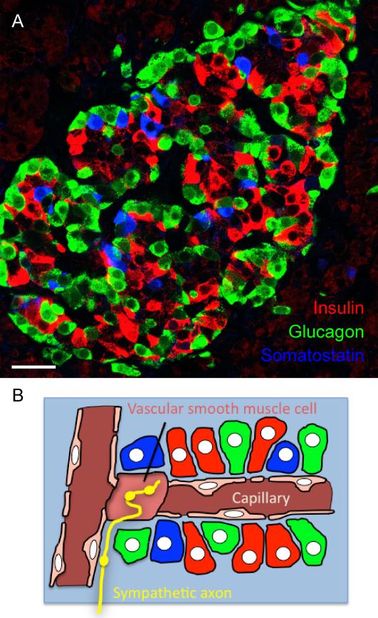
Figure 1. Cellular composition of pancreatic islets.
A, Confocal image of a human pancreatic section containing an islet immunostained for insulin (red, beta cells), glucagon (green, alpha cells), and somatostatin (blue, delta cells). These endocrine cells are distributed throughout the islet. Scale bar = 20 μm. B, Schematic diagram depicting endocrine cells (colors as in A), vascular cells (pink), and sympathetic axons (yellow) in the human islet. The vasculature in the human islet possesses numerous vascular smooth muscle cells embedded deep within the islet. These vascular cells are the main targets for sympathetic axons. The endocrine cells are aligned along the vessel without apparent order.

