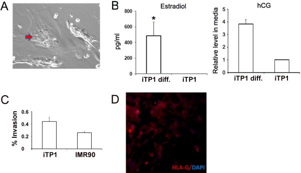
Fig4.
iTP cells can differentiate into trophoblast subtypes. (A) Formation of multi-nucleated trophoblast cells (red arrow). (B) Estradiol and hCG levels detected from day-7 culture medium. (n=3). (C) iTP cells showed higher % of invasion compared with donor fibroblasts (n=3). (D) Invasive cells were stained with HLA-G (Red) with Hochest 33342 fluorescence as nuclear counterstaining (blue). The original picture is 200×.

