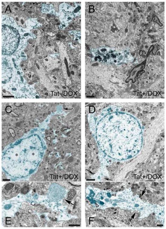
Figure 1.
Effects of Tat induction on the ultrastructure of astrocytes in CA1 region of the hippocampus (2-3-mo old mice). Astrocytes (highlighted in blue) from (A) control (Tat−/DOX) and (B-F) inducible, doxycycline (DOX) exposed Tat transgenic (Tat+/DOX) mice. (A) Astroglia in Tat−/DOX mice appear normal, displaying little or no pathology compared to astrocytes from Tat+/DOX animals. Grey matter astrocytes are distinguished from neurons by their lighter cytoplasm, are bordered by an irregular plasma membranes, lack organized cisternae of rough and large numbers of free ribosomes, lack microtubule bundles, possess more watery cytoplasm, and lack of synaptic contacts. (B-F) Astroglia in Tat+/DOX mice display increased numbers of inclusions with the features of lysosomes, autophagic vacuoles, and lamellar bodies compared to Tat−/DOX mice. In addition, the perikaryon above the astrocytic processes is that of a neuron (B, *). Note the abundance of endoplasmic reticulum-associated ribosomal clusters (Nissl bodies) and higher density of free ribosomes in the cytoplasm. (C) Whorls of membrane were occasionally seen within distal astrocyte processes (black arrow) shown at higher magnification in (E). Intranuclear vacuoles were present within the astrocyte cell body (D, *). (D) In the astrocyte highlighted in blue note the paucity of free ribosomes in the cytoplasm and very sparse rough endoplasmic reticulum (*). The black arrows denote the distal process of this astrocyte shown at higher magnification in (F). Numerous electron dense inclusions (white arrowheads) and vacuoles are present in this process. Scale bar for (A-D) = 1 μm, Scale bar for (E-F) = 0.5 μm.

