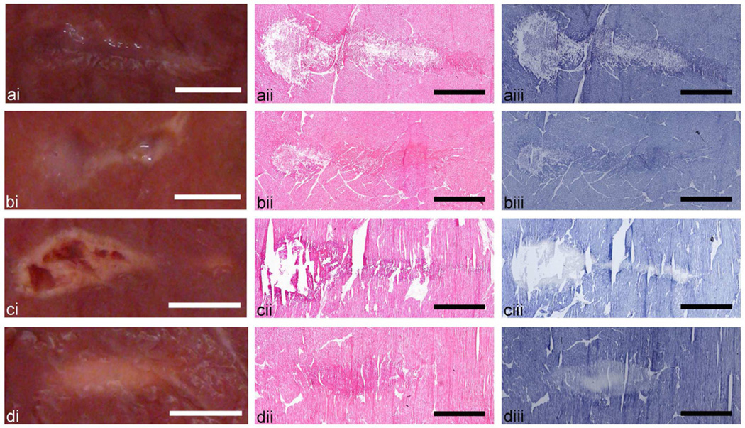
Figure 3.
Axial cross sections of the four types of HIFU lesions created in ex vivo bovine cardiac tissue: (a) liquid, (b) paste, (c) vacuolated thermal, and (d) solid thermal. Each lesion type is illustrated by (i) a representative photograph and histological images of consecutive frozen sections stained with (ii) H&E, revealing cellular structure, or (iii) NADH-d, revealing the extent of thermal damage. Lesions resulting from HIFU exposures that induce rapid boiling (a–c) are evident in both H&E and NADH stained sections as disrupted tissue. Thermal damage is indicated by the absence of dark blue NADH-d stain or by more eosinophilic (stained darker pink) areas in H&E stained sections (c, d) due to desiccation of cells. The NADH-d stained sections of neither liquid (a) nor paste (b) lesions show any indication of thermal damage. Scale bar represents 2 mm.

