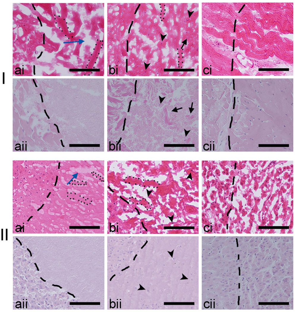
Figure 7.
Comparison of the H&E stained sections of fresh frozen (i) and formalin fixed (ii) lesions induced in bovine cardiac (I) and liver (II) tissue. The border of the lesions (to the right of images) is shown as the dashed line. Within the fresh frozen liquid (a) and paste (b) lesions in both tissues ice crystal formation was evident and resulted in voids (blue arrow) known as the freezing artifact, that is absent in the formalin fixed tissue. Within the paste lesion the intact nuclei (black arrow heads) and un-liquefied tissue fragments (black arrows) are better visualized in the formalin fixed sample (bii), although are discernible in the fresh frozen sample as well (bi). Fresh frozen and formalin fixed solid thermal lesions (c) are shown for comparison. Scale bar represents 100 µm.

