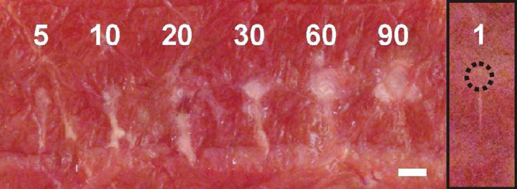
Figure 9.
A series of lesions induced in ex vivo bovine cardiac muscle using an increasing (5 – 90) number of 10-millisecond HIFU pulses delivered at a 1 Hz pulse repetition frequency. The corresponding in situ HIFU waveform is shown in Fig.1a. The number of pulses did not change the the type (“paste”) and the shape (“tadpole”) of the lesions. However, it affected their maximum width and length. Both of the lesion dimensions increased noticeably with the number of pulses up to 30, then reached saturation and virtually did not change starting from 60 pulses. The inset on the right shows the lesion resulting from a single pulse. The small circular void (outlined by a dotted line) at the anterior side of the upper lesion demonstrates, that even a single HIFU pulse, that induces millisecond boiling, can produce tissue emulsification. The scale bar is 2 mm.

