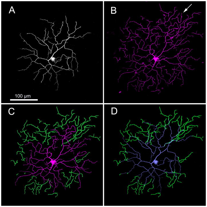
Figure 1.
Neurobiotin-staining of one type of rabbit ganglion cell reveals a bistratified dendritic tree with recurrent dendrites. (A) Focus on sublamina a shows that one set of dendrites ascends to and arborizes in the OFF sublamina. (B) Focus on sublamina b shows a second set of dendrites that arborize in the ON sublamina. However, a number of terminal dendrites can be seen in sublamina b that are not visibly connected to the soma in these layers (e.g., arrow). (C) Juxtaposition of the disconnected dendrites in sublamina b (green) highlights that they are not continuous with the remaining ON dendrites (red). (D) Juxtaposition of the disconnected dendrites in sublamina b (green) with the OFF dendritic arbor (gray) reveals that these dendrites originate from rapidly descending processes that originally arborized in sublamina a. These micrographs are stacks of 16 × 1 μm optical sections. This cell was 5.2 mm ventral to the visual streak. Increasing the gain of the green dendrites for emphasis produced a slight thickening of the dendrites compared to the other dendrites.

