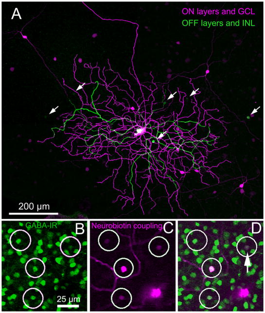
Figure 5.
(A) A Neurobiotin-injected ON bistratified ganglion cell is color-coded for depth (magenta is proximal; green is distal). Several amacrine cells with large somas (magenta) can be seen to have their somas displaced to the ganglion cell layer. Somas of a smaller amacrine cell type (green) are conventionally placed in the inner nuclear layer. (B–D) Amacrine cells seen after Neurobiotin-injection into another ON bistratified ganglion cell were colabeled with an antibody to GABA. (B) GABA-immunoreactive amacrine cells within a small region of the dendritic field. The larger magenta soma belongs to the injected ganglion cell. (C) Four coupled amacrine cells are shown. (D) Colocalization of Neurobiotin (magenta) and anti-GABA (green) signals. Somas of 3 of the amacrine cells were displaced to the GCL; the other was in the amacrine cell layer (arrow). Micrographs are stacks of (A) 25 × 1 μm and (B–D) 79 × 0.3 μm optical sections.

