
Figure 6.
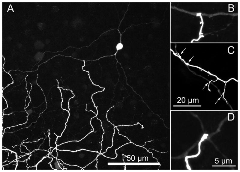
(A) The large displaced amacrine cells have a characteristic morphology with two dendrites emerging from opposite ends of soma and running 10–25 μm before branching. (B–D) Terminal varicosities from dendrites of ON bistratified ganglion cells frequently terminate on a dendrite of one of the coupled amacrine cells. The amacrine cell in B is a magnified view of the amacrine cell in A. Micrographs are stacks of (A) 22 × 1 μm (B,D) 1 × 0.3 μm (c) 3 × 0.3 μm optical sections.