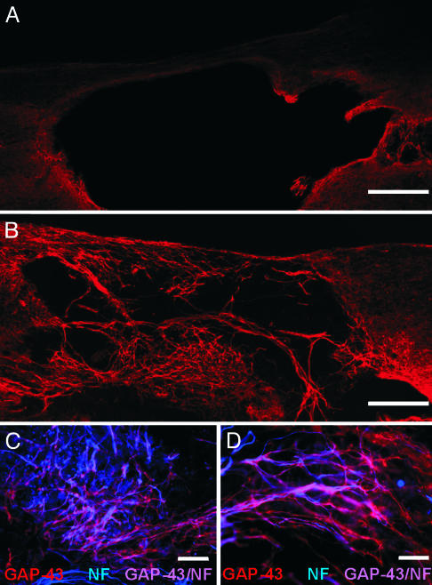
Fig. 1.
Implantation of fetal rat OECs is associated with a robust growth of GAP-43 immunopositive processes. (A) GAP-43 immunoreactivity around the perimeter of the cystic cavity was sparse in rats that received vehicle injections. (B) Many GAP-43 immunopositive fibers were observed extending into the cystic cavity in animals that received intraspinal implantation of OECs. Low (C) and high (D) magnification photomicrographs illustrating that many of the GAP-43 immunopositive fibers (red) coexpressed neurofilament (NF; blue). (Scale bars: 400 μm, A and B; 100 μm, C; 50 μm, D.)

