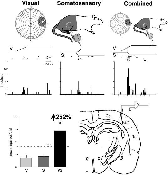
Fig. 2.
Receptive field overlap and multisensory enhancement in a visual-somatosensory neuron recorded at the occipital/parietal border. (Top) Visual and somatosensory receptive fields (shading) and locations of stimuli (icons depict stimulus movement) used in sensory testing. (Middle) Rasters and peristimulus time histograms illustrate responses to the visual, somatosensory, and combined visual-somatosensory stimulation. (Bottom Left) Summary bar graph illustrates the modality-specific [i.e., visual (V) and somatosensory (S)] and multisensory (i.e., VS) responses and the proportionate gain seen for the multisensory combination. (Bottom Right) The location of this neuron at the occipital/parietal border is shown on this drawing of a coronal section. *, P < 0.01. N, nasal; T, temporal; S, superior; I, inferior; Oc, occipital; Par1, parietal 1; Te, temporal.

