Figure 3.
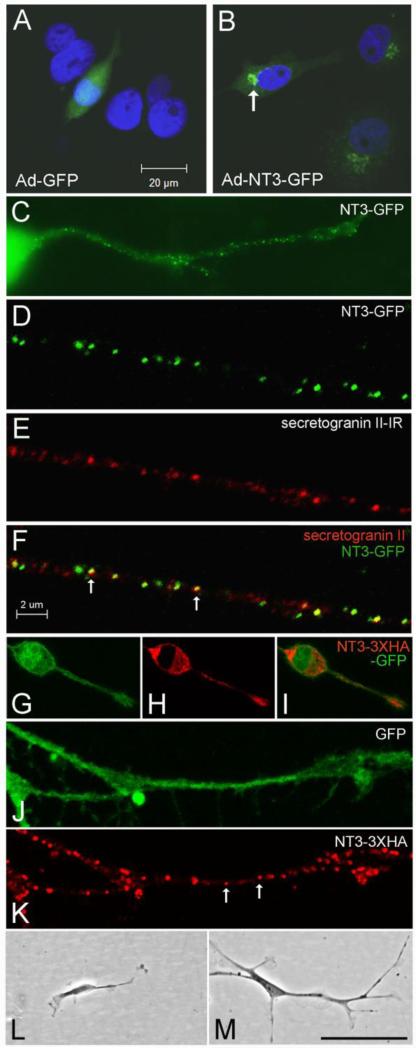
In vitro localization and bioactivity of NT3 fusion proteins. (A-B) Localization of GFP in COS cells infected with Ad-GFP (A) or Ad-NT3-GFP (B). The arrow in B indicates the fusion protein concentrated in the Golgi apparatus. (C) N2a cell infected with Ad-NT3-GFP showing punctate distribution of GFP within a neurite. (D-F) Confocal images illustrating co-localization of NT3-GFP with secretogranin II-IR (arrows in F) in a neurite extending from a cultured N2a cell. (G-K) N2a cells infected with NT3-3xHA-IRES-GFP contain GFP throughout (G), and HA-IR concentrated in the secretory apparatus, including a vesicular-like distribution within neurites (arrows in K). (L-M) Comparison of DRG neuron morphology at 4 days after treatment with conditioned medium from COS cells infected with Ad-GFP (L) or with Ad-NT3-3xHA-IRES-GFP (M). Bar in A= 20 μm for A-B. Bar in F= 2 μm for D-F, J-K. Bar in M= 20 μm in C, 42 μm in G-I, 100 μm in L-M.

