Figure 4.
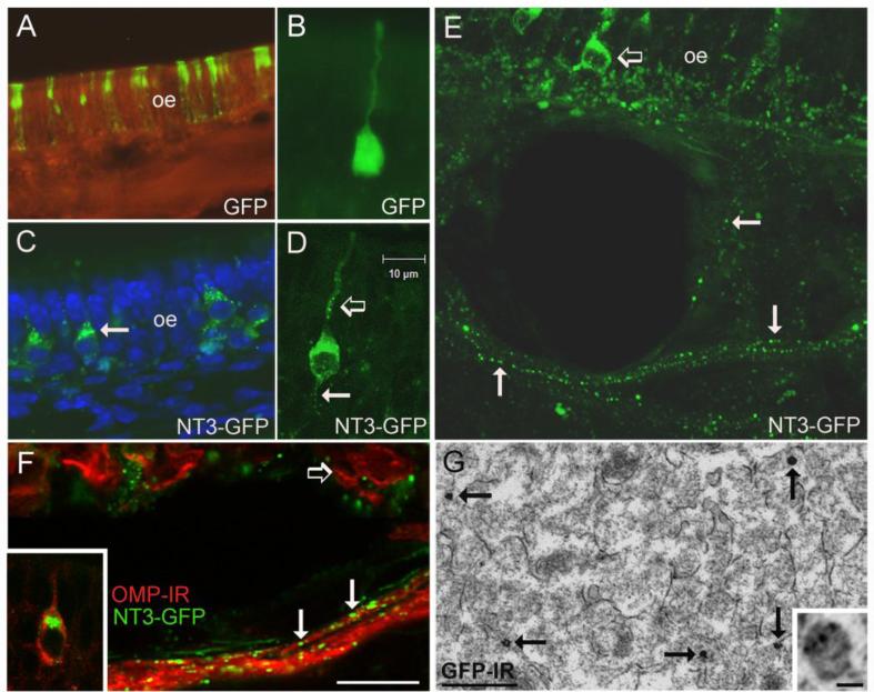
In vivo infection of olfactory sensory neurons. (A) Expression of GFP in a portion of the olfactory epithelium (oe) at 5 days after irrigation with the Ad-GFP control virus. The GFP label indicates both sensory neurons and sustentacular cells are infected. (B) An olfactory sensory neuron infected with Ad-GFP showing diffuse fluorescence throughout the cell. (C) Distribution of GFP in sensory neurons expressing NT3-GFP. The arrow indicates fluorescence concentrated in the Golgi apparatus. (D) A sensory neuron expressing NT3-GFP at 5 days after infection. Note the punctate distribution of GFP in the dendrite (open arrow) and in the proximal axon segment (arrow). (E) The vesicular-like distribution of NT3-GFP (arrows) can be followed in sensory axons exiting the epithelium. The open arrow indicates a GFP+ sensory neuron. (F) NT3-GFP+ puncta (arrows) are associated with olfactory sensory axons immunoreactive for OMP (red). The open arrow indicates OMP+ neurons in the epithelium, and the inset illustrates NT3-GFP concentrated in the Golgi apparatus of a mature, OMP+ sensory neuron. (G) Electron micrograph showing the distribution of GFP-immunogold-silver labeling in sensory axons in the olfactory nerve layer of a mouse treated with Ad-NT3-GFP. The arrows indicate dense gold-silver deposits, and the inset shows smaller deposits associated with a presumptive secretory vesicle that is less heavily labeled. Bar F= 58μm in A, 15μm in C, 20μm in E, 10μm in F. Bar in D=10μm in B and D. Bar in G= 500nm, inset=40nm.

