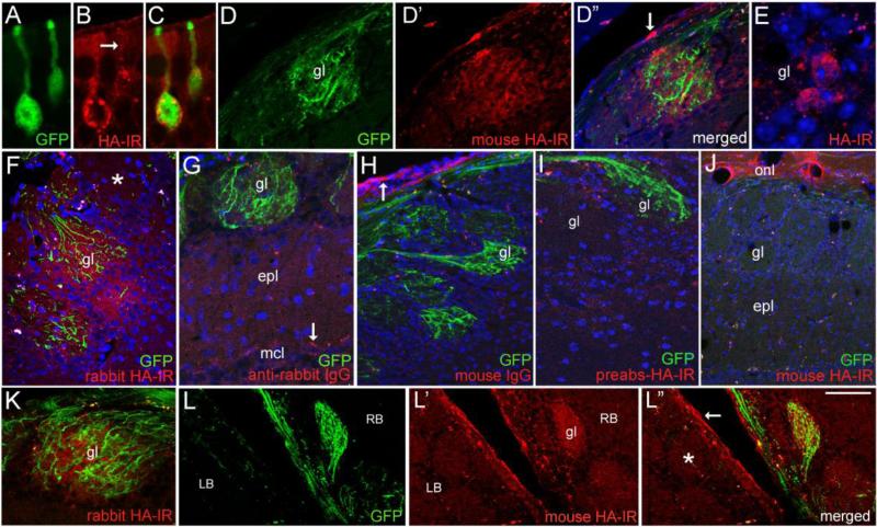
Figure 6.
Expression and distribution of NT3-3xHA in vivo. (A-C) Confocal images comparing the distribution of GFP and mouse HA-IR in sensory neurons 5 days post-infection (d.p.i.) with Ad-NT3-3xHA-IRES-GFP. Vesicular-like distribution of HA is seen in the dendrite (arrow). (D-D”) HA-IR is detected in a glomerulus (gl) containing GFP+ axons. The arrow in D” indicates non-specific labeling of ensheathing glia in the outer bulb nerve layer. (E) HA-IR in cell bodies bordering a glomerulus. HA-negative cells are present as well. (F) Glomerular staining produced by rabbit anti-HA antibody (Cell Signaling) at 7 d.p.i. The asterisk indicates a nearby glomerulus that lacks both GFP+ fibers and HA-IR. (G-I) Staining controls showing lack of HA-IR in sections processed without the rabbit primary antibody (G), incubated in mouse IgG (H), or treated with preabsorbed mouse anti-HA (I). The arrow in G indicates autofluorescent particles in mitral cells, and the arrow in H indicates non-specific labeling of ensheathing glia and processes by mouse IgG. (J) A section from the mouse shown in D, showing a bulb area lacking GFP+ axons. Glomerular HA-IR is also lacking, but non-specific staining of ensheathing glia processes occurs in the olfactory nerve layer (onl). (K) A GFP+/HA+ glomerulus at 7 d.p.i (6.5μm optical section thickness). (L-L”) Low magnification image of a horizontal section showing the medial right (RB) and left bulbs (LB) at 7 d.p.i. A GFP+ glomerulus (gl) in the right bulb is also HA+. GFP and HA-IR are lacking in glomeruli in the left bulb (asterisk). Non-specific staining occurs in the outer nerve layer (arrow in L”). epl, external plexiform layer; mcl, mitral cell layer. Bar in L”= 12μm in A-C and E, 40μm in D-D” and G, 38 μm in F and I, 64 μm in H, 80μm in J and L-L”, and 34 μm in K.

