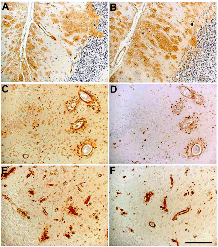
Fig. 3.
AB77 and AB76-2 immunoreactivity in FBD and FDD patients. Abundant AB77 (A) and AB76-2 (B) positive extracellular parenchymal deposits are seen in the cerebellum of a FBD patient. In addition, prominent vascular staining was detected in the dentate gyrus (C,D) and the CA4 region of the hippocampus (E,F) in a patient suffering from FDD. AB77: A, C, E; AB76-2: B, D, F. Scale bar: 100 µm

