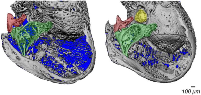
Figure 4.

Three dimensional micro-CT reconstructions of the whole ear bulbs from P3H1−/− and P3H1+/− littermates at P9. Middle ear bones are color coded as in Fig. 3 (malleus is green, incus is red, and stapes is yellow). The stapes is not identified in the P3H1 null ear due to the insufficient bone calcification.
