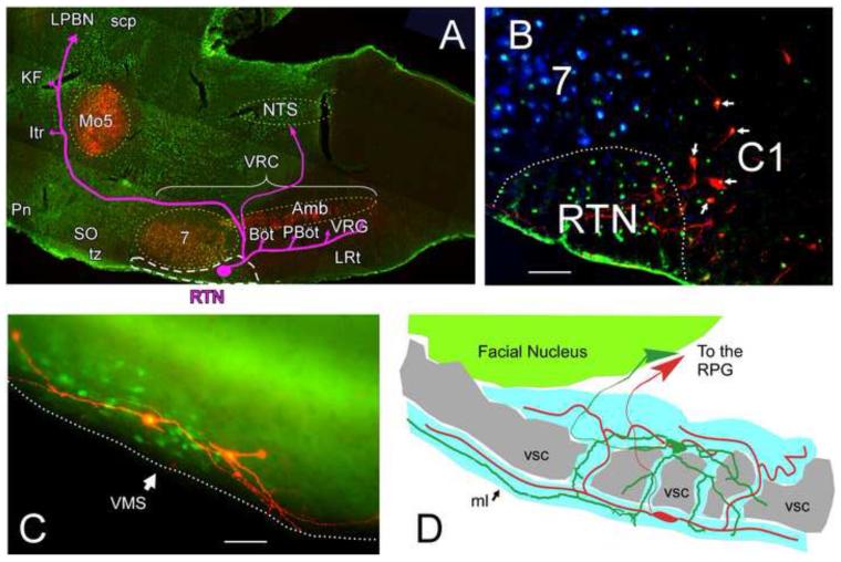
Figure 1.
Location of the Retrotrapezoid nucleus (RTN) and characteristic morphology of RTN neurons
A. Parasagittal section of rat brain. Phox2b–immunoreactive (ir) neuronal nuclei in green and choline acetyltransferase-ir (cholinergic) neurons in red. The RTN is outlined by a white dashed line and its known projections (Bochorishvili et al., 2011) are represented by the pink arrows. Other cell groups are indicated with yellow dashed outlines for anatomical reference. Abbreviations: Amb, nucleus ambiguus; Böt, Bötzinger Complex; Itr, inter trigeminal region; KF, Kölliker-Fuse; LPBN, lateral parabrachial nucleus; LRt, lateral reticular nucleus; Mo5, trigeminal motor nucleus; NTS, nucleus of the solitary tract; PBöt; Pre-Bötzinger Complex; Pn, pontine reticular nucleus; scp, superior cerebellar peduncle; SO, superior olive; tz, trapezoid body; VRG, ventral respiratory group.
B. Transverse section of rat brain through the rostral medulla oblongata showing the ventral region centered about 2 mm lateral to the midline (midline is to the right) with tyrosine hydroxylase-ir neurons in red (C1), facial motor neurons in blue and Phox2b-ir neurons in green (unpublished illustration from (Stornetta et al., 2006)). Note that the C1 neurons also show Phox2b-ir nuclei (appearing yellow, arrows). The RTN is indicated by the dashed white outline. 7, facial motor nucleus. Scale bar: 50 m
C. pH-sensitive RTN neurons filled with biotinamide (red) during whole cell recording. These neurons were recorded in tissue from a Phox2b-EGFP transgenic mouse in which expression of the fluorescent protein (green) was limited to a subset of Phox2b-expressing neurons that included the RTN (modified from (Lazarenko et al., 2009)). Scale bar: 25 m.
D. Schematic drawing of two RTN chemosensitive neurons recorded in a halothane-anesthetized adult Sprague-Dawley rat. These neurons, juxtacellularly labeled with biotinamide and reconstructed through adjacent sections with the aid of a computer driven stage and Neurolucida software (Mulkey et al., 2004), are representative of the population at large. The cell bodies of RTN neurons reside within the region outlined in blue which includes the marginal layer (ml; red cell) and bands of neuropil that are interspersed between the myelinated axon bundles of the ventral cerebellospinal tract (vsc; in gray) and include the more dorsally located green cell. Regardless of the location of their cell bodies, RTN neurons have at least one long dendrite running very close to and parallel to the ventral surface of the brain within the marginal layer. RPG: respiratory rhythm and pattern generator, a network of interconnected neurons located in Böt, pBöt, VRG, KF, Itr and NTS as defined in panel A.

