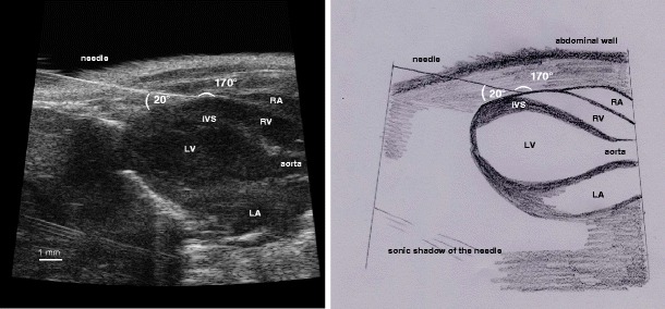
Fig. 2.

Echocardiographic visualisation of pericardial injection: modified echocardiographic LAV showing the position of the needle tip inside the pericardial sac for the pericardial injection in characteristic RV position (left) and scheme (right). IVS interventricular septum, LA left atrium, LV left ventricle, RA right atrium, RV right ventricle
