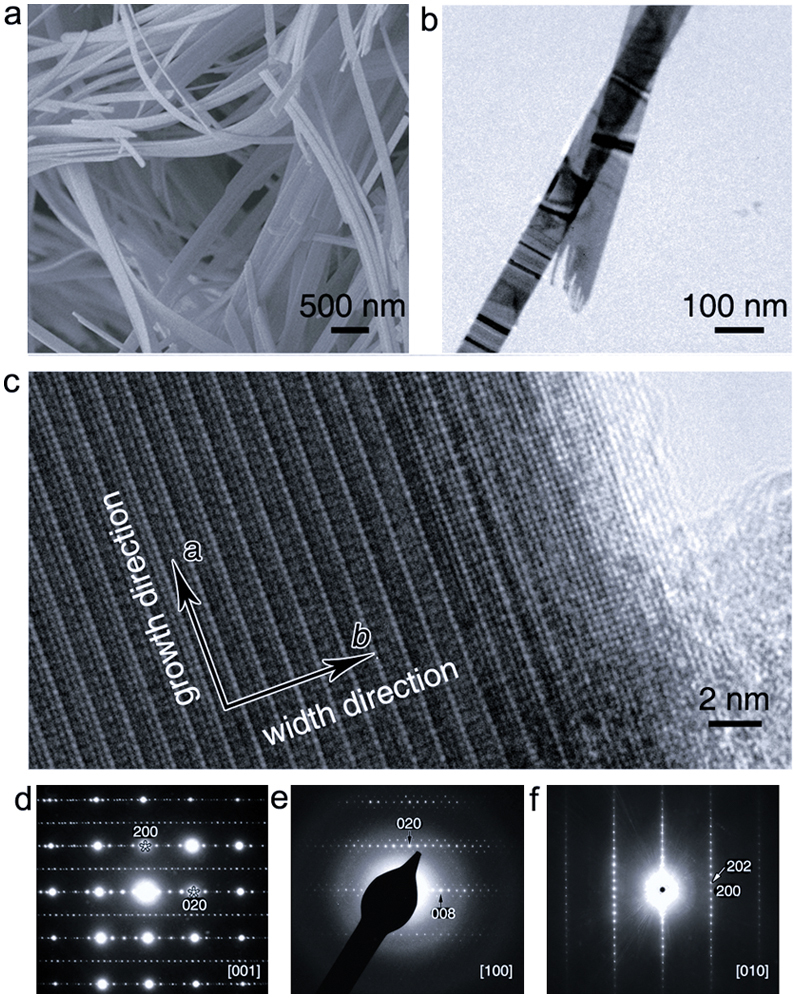
Figure 2. Morphology characterizations of the Bi4.2K0.8Fe2O9+δ nanobelts.

(a–c) Scanning electron microscopy (a), bright-field transmission electron microscopy (b), and [001] zone axis high-resolution transmission electron microscopy (c) images of the nanobelts. (d–f), [001] (d), [100] (e), and [010] (f) zone axes selected area electron diffraction patterns (SAED) of the nanobelts, and a commensurately modulated wave vector q* = (0, 0.25, 1) can be confirmed.
