
Figure 1.
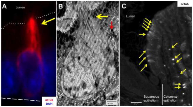
A) Gastric tuft cell identified by microtubule staining (yellow arrow) with anti-acetylated-α-Tubulin (acTub, red). The dotted line indicates the apical border, and the dashed line the basal membrane. B) Electromicrograph of a tuft cell in the stomach, showing the characteristic narrow apex with blunt microvilli and numerous microtubules (yellow arrow) that extend along the cell body, and lateral branches (red arrow). C) Tuft cells identified with acTub (yellow arrows) were found in high numbers at the squamous/columnar epithelium border of the stomach.