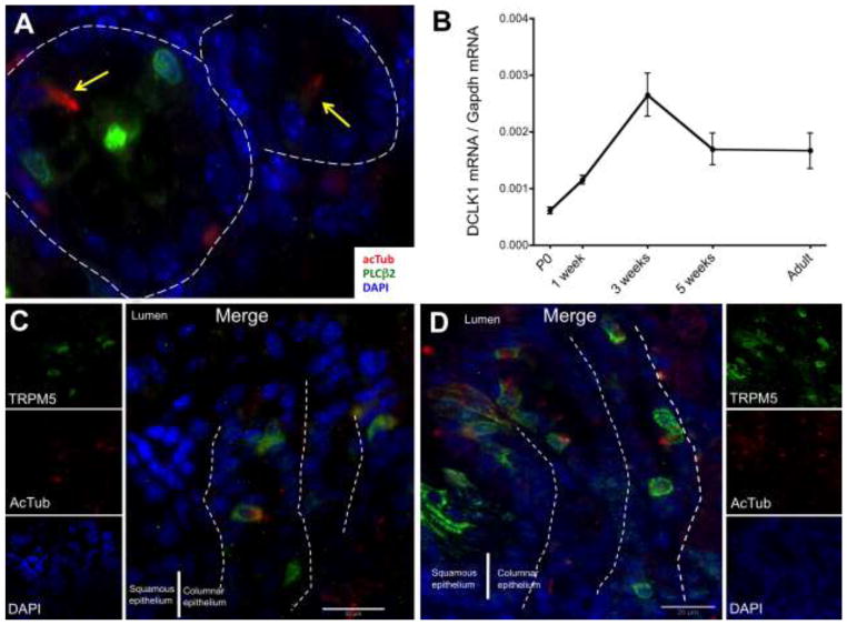
Figure 4.
A) In the embryonic stomach (E18.5), tuft cells (yellow arrows) were not positive for PLCβ2 (green). In the gastric corpus, DCLK1 mRNA abundance was increased after birth (B) and tuft cells were found in low numbers by 2 weeks (C). The high number of tuft cells in the squamous/columnar epithelium junction observed in the adult was reached only after 3 weeks of age (D). The epithelial basal membrane is marked with white dashed lines.

