Figure 5.
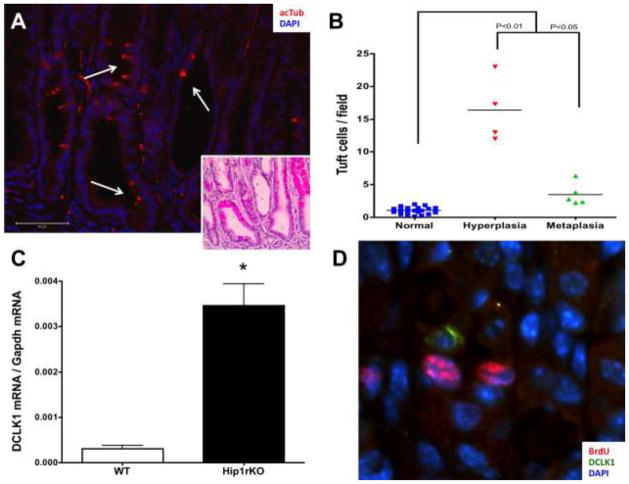
Tuft cells are expanded in hyperplasia in the stomach. A) A representative image of a hyperplastic area in the corpus of an 11 month-old gastrin knockout mouse (GKO). Tuft cells are identified with acTub (red, white arrows), and the hematoxylin-eosin stain of the same area is shown in the insert. B) Quantification of tuft cells observed in GKO mice corpus. GKO mice develop antral and corpus hyperplasia and corpus metaplasia. Areas in which expanded expression of spasmolytic peptide were observed were considered metaplastic. Tuft cells in the surrounding normal mucosa in the same sample were quantitated. P values were calculated using One-way ANOVA with Dunnet’s post-test. C) Increased expression of DCLK1 was also observed in 5 week-old Hip1rKO mice, which develop SPEM and corpus hyperplasia. Data presented are mean ±S.E.M. (N = 3; *P<0.005 with Student’s T-test). D) DCLK1 positive cells (green) were not proliferating in Hip1rKO 3 week old mouse corpus (BrdU staining in red).

