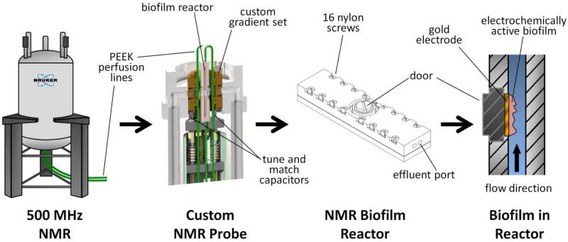
Figure 1. Nuclear magnetic resonance microimaging system for studying EABs.
An illustration of the experimental arrangement for NMR used to study diffusion in EABs. The diagrams show (from left to right): the vertical bore superconducting magnet with perfusion lines leading to the bottom-loaded NMR probe (medium flowing against gravity); a cutaway view of the custom NMR probe, shown holding the NMR biofilm reactor; an external view of the NMR biofilm reactor; and a cutaway view of the NMR biofilm reactor containing a perfused EAB growing on a gold disc electrode.

