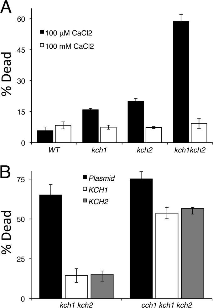
Fig 2.
Kch1 and Kch2 are essential for maintaining cell viability. (A) The strains listed in Fig. 1C were exposed to 25 μM α-factor in SC-100 media with or without additional 100 mM CaCl2 and stained with propidium iodide. Live and dead cells were counted by flow cytometry. (B) Strains kch1 kch2 and cch1 kch1 kch2 (CS03 and CS07) were transformed with a control plasmid or plasmids that overexpress the KCH2-HA3 or KCH1-HA3 genes (pSM10, pCS01, and pCS02), exposed to 50 μM α-factor, and then analyzed for pheromone-induced cell death as in panel A. The averages of three biological replicates (± the SD) are shown.

