Fig 7.
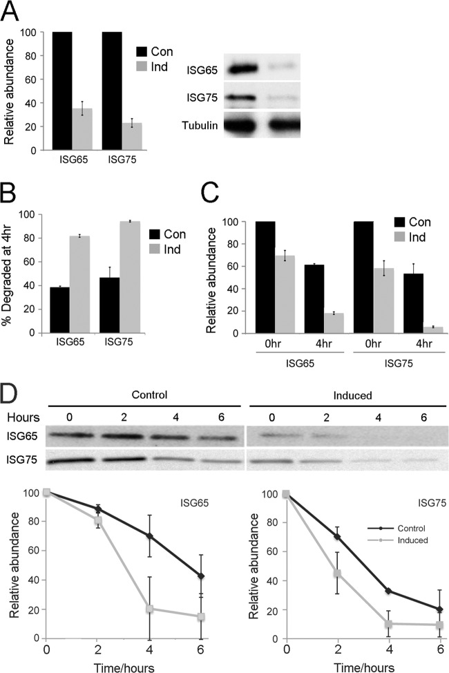
Influence of RME-8 on ISG65 and ISG75 stability. (A) Expression of ISG65 and ISG75 in RME-8 RNAi cells 24 h postinduction of TbRME-8 knockdown detected by Western blotting. Steady-state ISG expression is decreased by approximately 3-fold compared to control cells as assessed in three independent experiments and normalized to tubulin expression. (B) ISG turnover was analyzed in radioimmunoprecipitation assays (RIPAs). Cells were pulse-labeled with [35S]methionine-cysteine for 1 h and chased for 4 h. In RME-8 RNAi cells 70% to 90% of labeled ISGs were degraded after a 4-h chase compared to 40% to 50% in control cells, indicating an accelerated ISG turnover as an effect of RME-8 depletion. (C) ISG biosynthesis as analyzed by 35S incorporation for 1 h in RIPAs was decreased to 70% for ISG65 and 60% for ISG75 24 h postinduction compared to control cells (0-h time point). After a 4-h chase (4-h time point), the induced RNAi lines showed much lower abundance of ISG65 and ISG75 than control cells, indicative of faster turnover, as shown in panel B. (D) ISG65 and ISG75 are destabilized after RNAi knockdown of TbRME-8 as seen by Western blotting of whole-cell lysates after treatment with cycloheximide. The ISG half-life was reduced from ∼4.5 h in control cells to ∼2.5 h in induced cells. Data represent the means of the results of two independent experiments normalized to the values for tubulin; standard deviations are indicated.

