Fig 4.
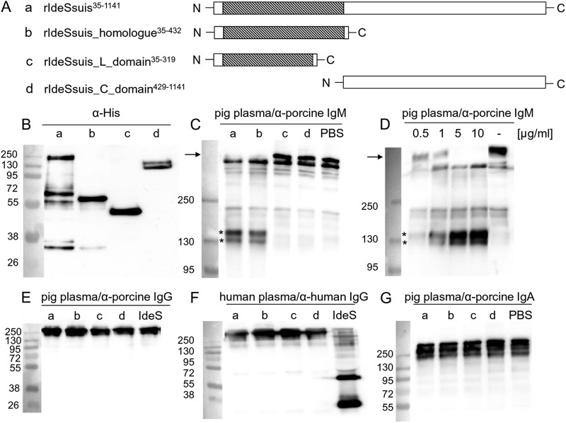
Cleavage of IgM but not IgG and IgA by recombinant IdeSsuis constructs. (A) Illustration of rIdeSsuis and its truncated derivatives. Regions similar to IdeS of S. pyogenes are shaded. (B) Anti-His Western blot analysis of purified rIdeSsuis (a) and its truncated derivatives (b to d) illustrated in panel A. (C) Anti-IgM Western blot (PAB) of diluted porcine plasma incubated with 5 μg/ml of rIdeSsuis (a) or its truncated derivatives (b to d) illustrated in panel A. (D) Anti-IgM Western blot analysis (PAB) of diluted porcine plasma incubated with increasing concentrations of rIdeSsuis as indicated or with PBS for 10 min at 37°C. (E, F) Anti-porcine (E) and anti-human (F) IgG Western blot analysis of diluted porcine and human plasma, respectively, incubated with 5 μg/ml of rIdeSsuis (a) or its truncated derivatives (b to d) shown in panel A. Recombinant IdeS of S. pyogenes was included as positive control for cleavage of human IgG. (G) Anti-porcine IgA Western blot analysis of diluted porcine plasma incubated with 5 μg/ml rIdeSsuis (a) or its truncated derivatives (b to d) illustrated in panel A. The final dilution of porcine plasma was 1:100 in all samples. The marker bands are shown on the left (sizes in kDa).

