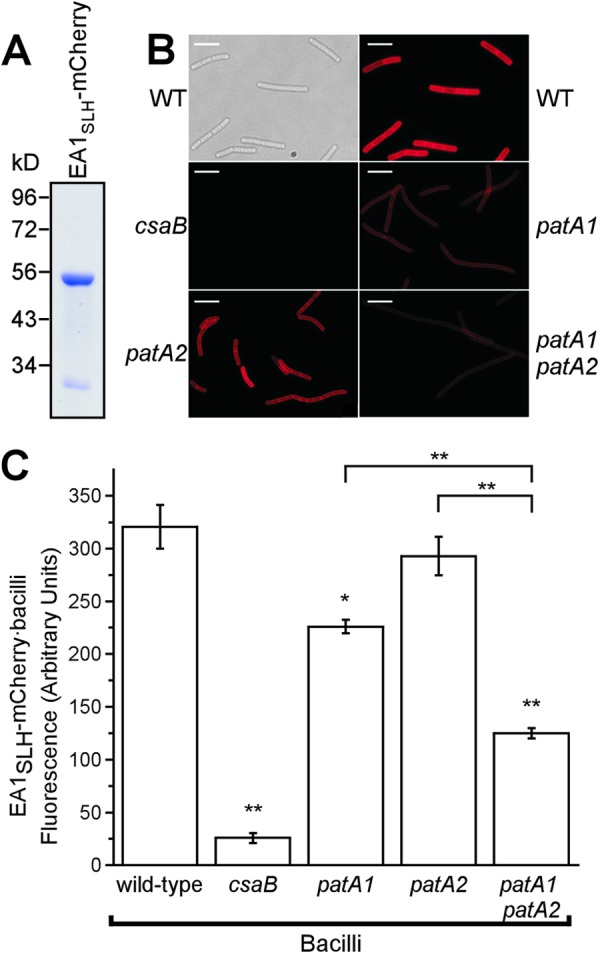
Fig 4.

The SLH domain of EA1 displays reduced binding to the SCWP from patA1 patA2 mutant B. anthracis. (A) Coomassie-stained SDS-PAGE gel of the purified fusion protein EA1SLH-mCherry. Numbers indicate the positions of molecular mass markers (in kDa). (B) Vegetative forms of B. anthracis strains were stripped of their S-layers via treatment with 3 M urea. Differential interference contrast (DIC) and fluorescence microscopy images of the wild type and fluorescence microscopy images of csaB, patA1, patA2, and patA1 patA2 mutant B. anthracis strains incubated with EA1SLH-mCherry are shown. Scale bar, 15 μm. (C) The fluorescence intensity of EA1SLH-mCherry binding to the envelope of S-layer-stripped B. anthracis strains was quantified in three independent experimental determinations. Data were averaged and examined with an unpaired two-tailed Student's t test. *, P < 0.01; **, P < 0.001.
