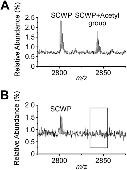
Fig 7.

Ion signals for acetylated SCWP in wild-type but not in patA1 patA2 mutant B. anthracis cells. (A) Ion signals from the wild-type SCWP were detected with MALDI-TOF mass spectrometry; m/z 2,800.7 was interpreted as the sodiated ion [ManNAc-GlcNAc2(Gal3)]2-ManNac-GlcNAc(Gal), and m/z 2,842.8 was interpreted as its acetylated variant. (B) Ion signals from the patA1 patA2 mutant SCWP were detected with MALDI-TOF mass spectrometry; m/z 2,800.7 was interpreted as the sodiated ion [ManNAc-GlcNAc2(Gal3)]2-ManNac-GlcNAc(Gal). m/z 2,842.7 was not detected in the spectrum. Of note, although mass spectrometry data are in agreement with the proposed compound structures, they do not constitute experimental proof for the structural assignments.
