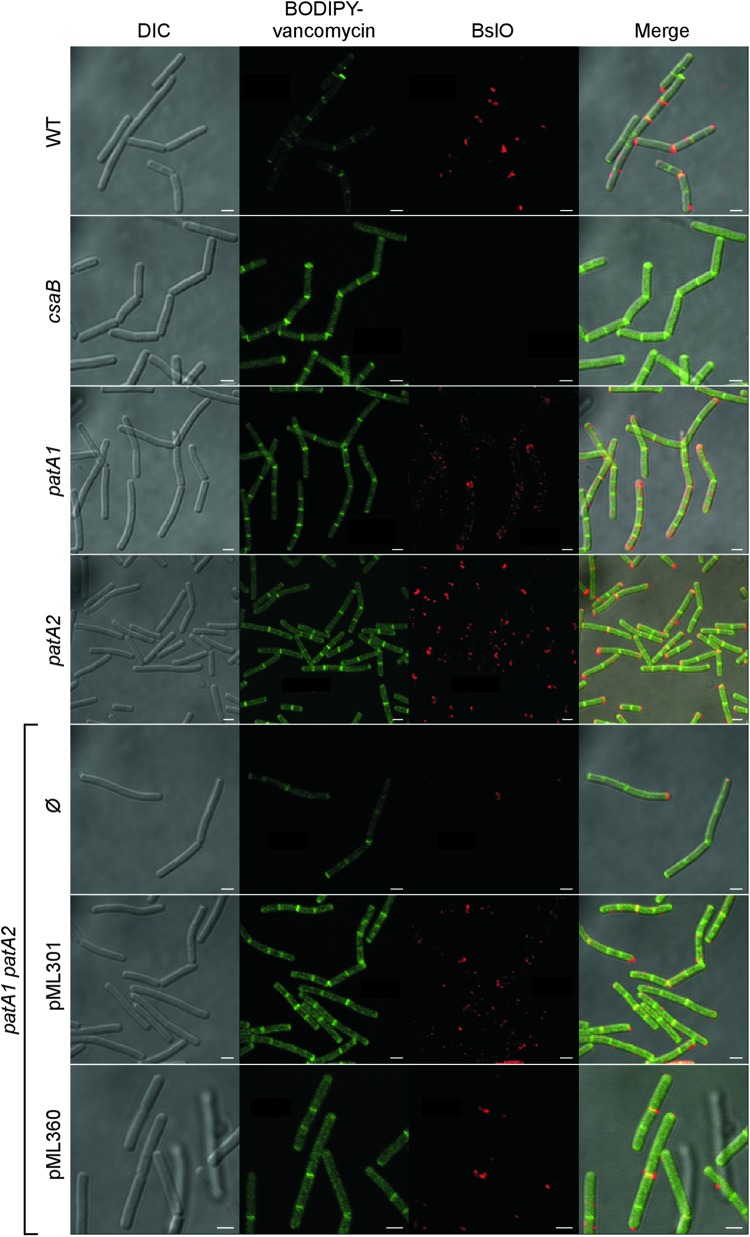
Fig 9.
Localization of the S-layer protein EA1 in the envelope of wild-type, csaB, patA1, patA2, and patA1 patA2 B. anthracis strains. Spores from wild-type and mutant B. anthracis strains were diluted into BHI broth, and germinated bacilli were incubated for 3 h. Vegetative forms were fixed in 4% buffered formalin. Differential interference contrast (DIC) and fluorescence microscopy with BODIPY-vancomycin or rabbit polyclonal antibodies staining against the S-layer-associated protein BslO followed by secondary antibody DyLight594 conjugate were used to acquire images. Data sets were merged to reveal the location of cell wall septa (BODIPY-vancomycin) and the S-layer-associated protein BslO in wild-type and mutant bacilli. The patA1 patA2 mutant strain was transformed with plasmid pML301, expressing patA1 and patB1, or pML360, expressing patA2 and patB2, or left untransformed (Ø). Scale bar, 1 μm.

