
Fig 3.
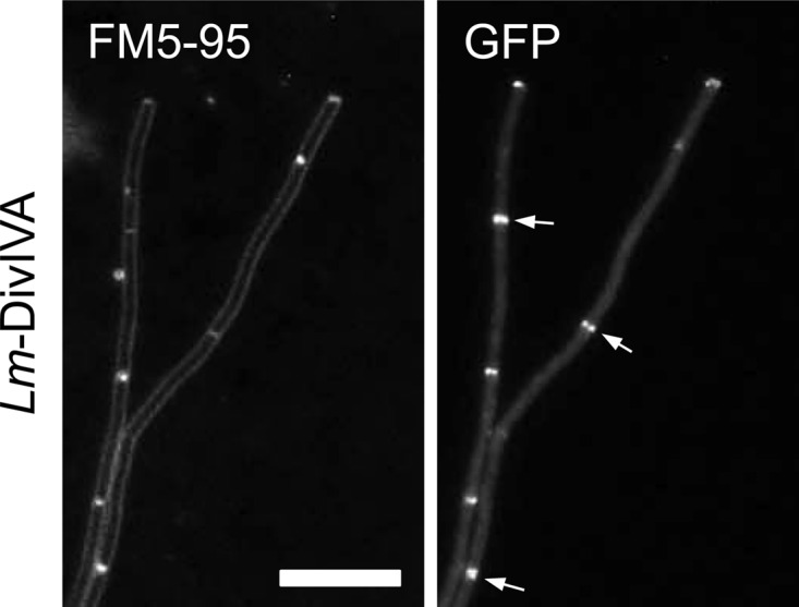
Localization of L. monocytogenes DivIVA-GFP in a B. subtilis ΔdivIVA background. Strain BSN373 (expressing L. monocytogenes DivIVA-GFPA206K) was grown in LB medium supplemented with 0.5% xylose. The localization pattern of L. monocytogenes DivIVA-GFP was analyzed by epifluorescence microscopy (right), and for orientation, a FM5-95-stained image (left) was taken in parallel. Several septal DivIVA-GFP signals are indicated by arrows. Bar, 5 μm.