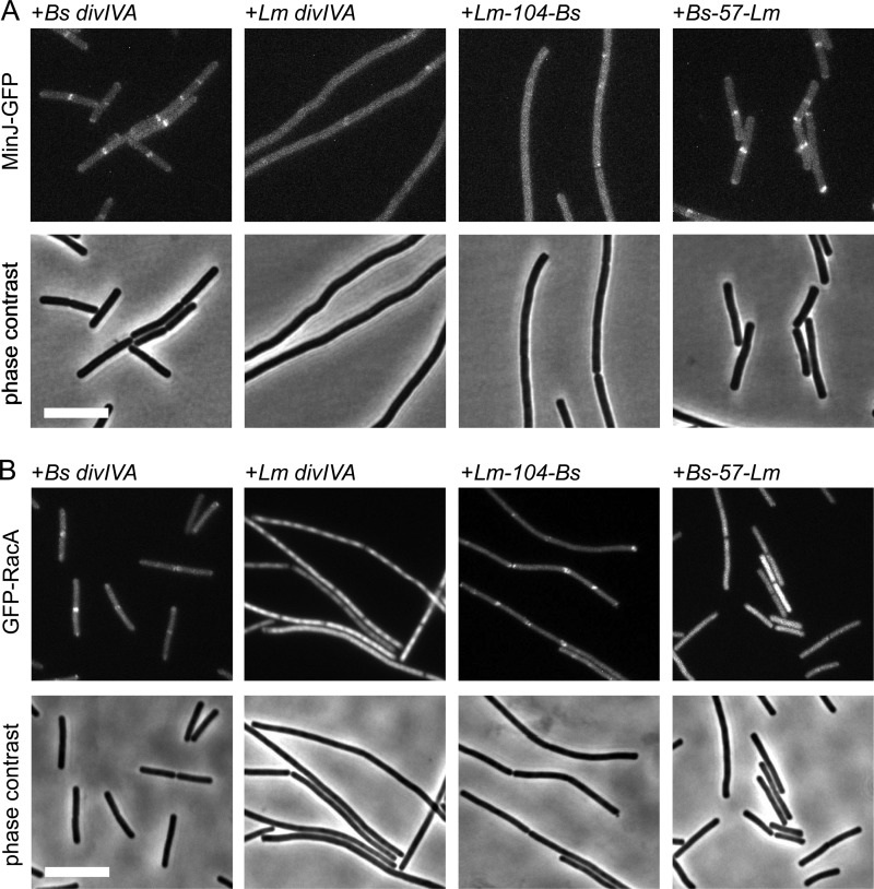
Fig 6.
Localization of MinJ and RacA in B. subtilis strains expressing selected L. monocytogenes and B. subtilis divIVA chimeras. (A) Fluorescence micrographs showing the subcellular localization of MinJ-GFP in L. monocytogenes and B. subtilis divIVA chimera strains during mid-logarithmic growth in LB broth supplemented with 0.5% xylose at 37°C (top). MinJ-GFP was imaged in strains expressing the Lm-104-Bs DivIVA (strain BSN336) and the Bs-57-Lm DivIVA (strain BSN338) chimeras. As a control, MinJ-GFP was also visualized in ΔdivIVA strains which express B. subtilis divIVA (strain BSN334) or L. monocytogenes divIVA (strain BSN335). Phase-contrast images were included for better orientation (bottom). (B) Localization of RacA in B. subtilis strains expressing the same L. monocytogenes and B. subtilis divIVA chimeras as in panel A. Fluorescence images were obtained on cells during growth in LB broth containing 0.5% xylose at 37°C (top). GFP-RacA was visualized in strains expressing the Lm-104-Bs DivIVA (strain BSN342) and Bs-57-Lm DivIVA (strain BSN344) chimeras. As controls, GFP-RacA was also imaged in strain BSN340, which expresses B. subtilis divIVA, and in strain BSN341, which expresses L. monocytogenes divIVA. Phase-contrast images were included for better orientation (bottom). Bar, 5 μm.

