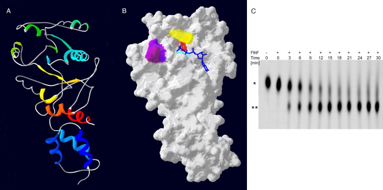
Fig 1.
FlhF of P. aeruginosa is a GTPase. (A) Amino acids 150 to 401 of FlhF were modeled by the Phyre2 server and are shown threaded onto the BsFlhF structure (PDB file 2PX0D). The ribbon diagram is colored by secondary structure succession (blue to red) and is shown in the same orientation as in panel B. (B) Molecular surface model of PaFlhF. Mutated residues are colored as follows: K222, aqua; R251, yellow; D294, red; L298, purple; and P299, dark plum. The positions of GTP (blue) and Mg2+ (green sphere) were predicted using 3DLigandSite. (C) Wild-type FlhF was incubated with 50 nM [α-32P]GTP. The reaction mixture was sampled at the indicated time points, and the reaction was stopped by the addition of 200 mM MgCl2. Reaction products were separated on TLC plates and visualized by autoradiography. The positions of GTP (*) and GDP (**) are indicated.

