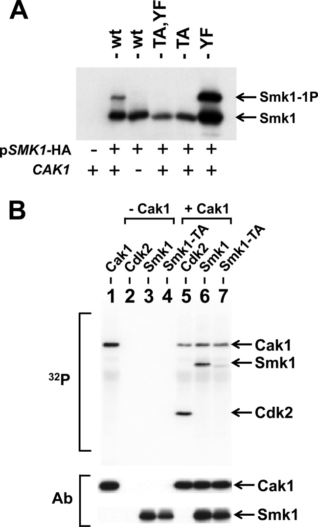
Fig 2.
Cak1 phosphorylates Smk1 on T207. (A) Smk1-HA was constitutively produced using the mseΔ promoter in cdc28-43244 (hyperactivated) CAK1 (+) and cak1Δ (−) mitotic cells, and proteins were analyzed by electrophoresis through Phos-tag gels and immunoblotting using an HA antibody. (B) Phosphotransferase assays containing the indicated proteins were carried out in the presence of 32P-labeled ATP. The proteins were resolved on electrophoretic gels and transferred to a membrane, and radioactivity was recorded using a phosphorimager (32P). Lane 1, Cak1-GST; lane 2, mammalian Cdk2; lane 3, Smk1 immunoprecipitated from an in vitro transcription/translation (TNT) reaction; lane 4, Smk1-T207A immunoprecipitated from a TNT reaction; lane 5, mammalian Cdk2 plus Cak1-GST; lane 6, immunoprecipitated Smk1 plus Cak1-GST; lane 7, immunoprecipitated Smk1-T207A plus Cak1-GST. The positions of the proteins are indicated by arrows. The same filter was subsequently assayed for Cak1 or HA (Smk1) immunoreactivity (Ab) as indicated. The incorporation of radioactivity into the Cak1 band requires the GST moiety since untagged Cak1 does not undergo autophosphorylation in these assays.

