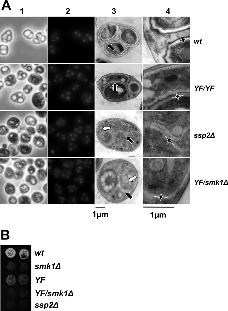
Fig 5.
The smk1-Y209F/smk1Δ phenotype is indistinguishable from the ssp2Δ/ssp2Δ phenotype. (A) Cells with the indicated genotypes were harvested 24 h after meiotic induction. Samples were stained with DAPI and images recorded using phase-contrast (column 1) and fluorescence (column 2) microscopy. Aliquots of these samples were stained with uranyl acetate and examined by thin-section electron microscopy. A representative low-magnification electron micrograph (column 3) and corresponding high-magnification electron micrograph (column 4) are shown. Each black arrow in column 3 points to the center of a magnified area shown in column 4 for reference. The elongated vertices of the stars in column 4 point to the inner layers of the spore wall. Note that in the wild type (wt), these inner layers are surrounded by the chitin/chitosan and the electron-dense dityrosine coat (outer layers). In the smk1-Y209F/smk1-Y209F (YF/YF) spores, the indicated inner layers (star) are surrounded by the chitin/chitosan layer and dityrosine coat resembling wild-type spore walls, while the inner layers surrounding the adjacent spore (indicated by the short upward-pointing vertex of the star) are absent. While the inner layers are present in ssp2Δ and smk1-Y209F/smk1Δ (YF/smk1Δ) spores, the outer layers are missing in almost all spores. The white arrows in the ssp2Δ and YF/smk1Δ samples in column 3 point to the supernumerary vesicles that appear to be surrounded by inner spore wall layers. (B) Sporulating cells of the indicated genotypes were harvested 24 h after meiotic induction in duplicate, equivalent numbers of cells were collected on filters, and the incorporation of dityrosine into insoluble material was assayed using the fluorescence assay.

