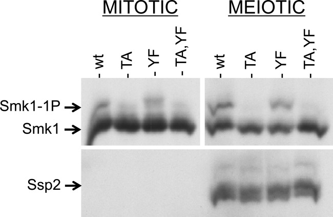
Fig 6.
Smk1 produced prior to meiotic induction is not autophosphorylated on Y209 in sporulating cells. Vegetative smk1Δ SSP2-MYC cells harboring plasmids expressing the indicated SMK1-HA mutant from the mseΔ promoter were transferred to sporulation medium. Samples were taken at 0 h and at 9.5 h and analyzed by electrophoresis through Phos-tag gels and immunoblotting using an HA (Smk1) or a MYC (Ssp2) antibody. There is more Smk1 in the mitotic cells (left) than in the meiotic cells (right) due to degradation of Smk1 that takes place during the 9.5-h interval. The mitotic and meiotic Smk1 signals were analyzed in the same blot, but the exposure of the meiotic samples was approximately 4 times as long as the mitotic exposure to facilitate the comparison of the relative levels of phosphorylated Smk1 in mitotic and meiotic cells. TA, T207A; YF, Y209F; wt, wild type.

