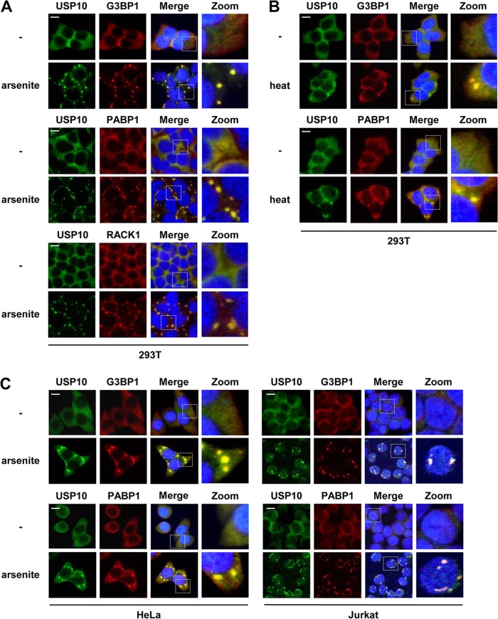
Fig 4.
Subcellular localization of USP10 and SG marker proteins. (A to C) 293T (A and B) and HeLa and Jurkat (C) cells were treated with 0.5 mM sodium arsenite for 60 min or heat shock at 42°C for 60 min as indicated. The cells were then fixed and stained with anti-USP10-C (green) together with anti-G3BP1 (red), anti-PABP1 (red), or anti-RACK1 (red). Nuclei were counterstained using Hoechst 33258 (blue). The bars indicate 10 μm.

