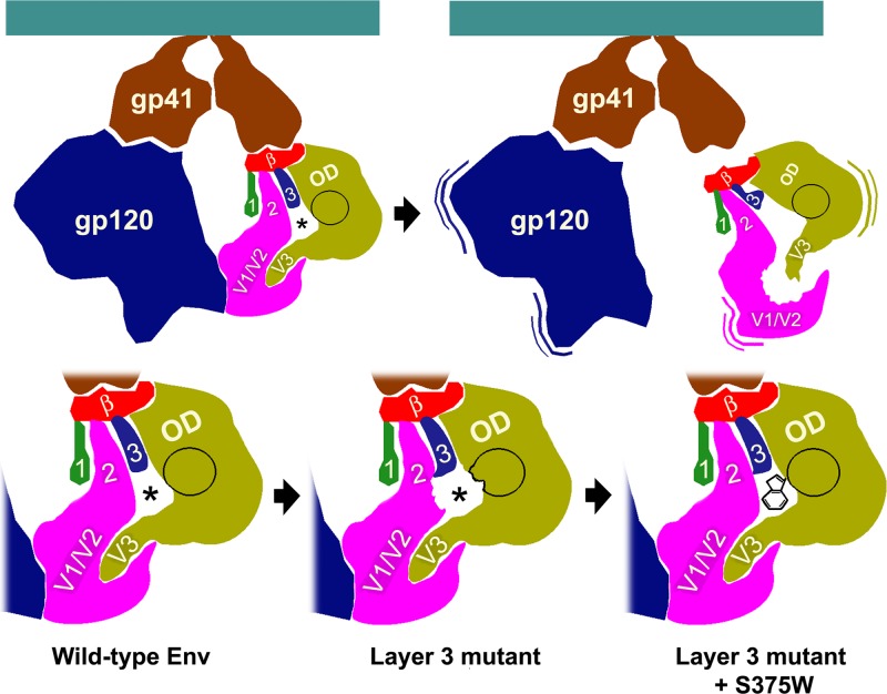
Fig 8.
Model for the role of layer 3 in HIV-1 Env trimer stability and CD4 binding. The image on the top left depicts the unliganded HIV-1 Env trimer, with two gp120 subunits and two gp41 subunits visible from the perspective shown. The gp120 subunit on the right is subdivided into the outer domain (OD), inner domain (β-sandwich [red] and layers 1, 2, and 3), and the TAD (V1/V2, and V3 regions) (35). The initial site of CD4 binding is circled in black, and the location of the Phe43 cavity is shown by an asterisk. Layer 3 modulates the interaction of the outer domain and layer 2 in the inner domain. The upper right image shows the consequences of changes in Trp479, which is located in layer 3, in the gp120 interdomain interface. Changes in Trp479 significantly disrupt the relationship of the inner and outer domains. Because the V1/V2 and V3 regions are anchored in the inner and outer domains, respectively, the gp120 TAD, which comprises the V1/V2 and V3 regions, is disrupted. Thus, changes in Trp479 lead to trimer instability and gp120 shedding. The images in the bottom row illustrate the phenotypic consequences of changes in layer 3 that are less disruptive than the alteration of Trp479. These more subtle changes in layer 3 cause local alterations of gp120 conformation around the Phe-43 cavity that affect CD4 binding, with effects on both on-rate and off-rate (bottom panels). The more subtle changes in layer 3 can be compensated by the S375W change, which fills the Phe43 cavity with the indole ring of tryptophan.

