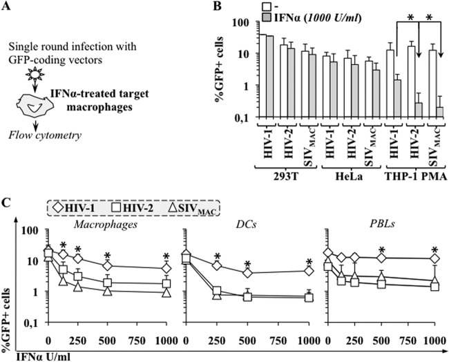
Fig 3.
Primate lentiviruses exhibit distinct susceptibilities to IFN-α during the early phases of infection. (A) Schematic representation of the assay used here. (B) Established cell lines were pretreated with IFN-α for 24 h then challenged with equal amounts of single-round infection-competent VSVg-pseudotyped CMV-GFP bearing HIV-1, HIV-2, and SIVmac vectors (MOI between 0.2 and 0.5). The percentage of GFP-positive cells was determined 3 days afterward using flow cytometry. (C) Macrophages, monocyte-derived dendritic cells (DCs), and PHA-activated PBLs were treated similarly (except that vectors were used at MOIs between 2 and 5) and analyzed. The panels present averages and SEMs obtained in 3 to 10 independent experiments, each of which was carried out with cells obtained from different donors. The asterisk indicates a P value of ≤0.05, according to an unpaired Student t test, between the defects observed upon treatment with IFN-α in HIV-1 versus HIV-2 (or SIVmac).

