
Fig 8.
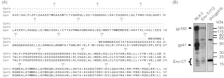
Sequence alignment of authentic Gp41 and D2b-derived isoforms Gp41a and Gp41b. From position 208, all of the protein isoforms show sequence identity. (B) Western blot analysis with Gp41-CT antibodies employing lysates of cells transfected or infected with the indicated constructs. The positions of molecular mass markers are shown on the right, and those of the provirally expressed Env-CT-containing proteins are shown on the left. The Env-CT bands are a group of specifically detected low-molecular-mass protein entities.