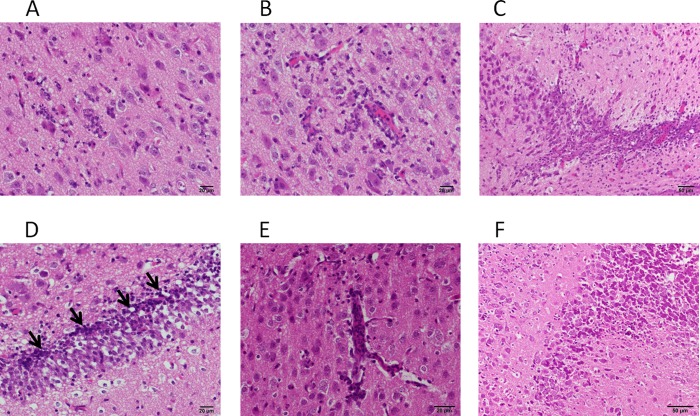
Fig 7.
Histopathological examination of the brains of infected hamsters. The sections from the brains of infected hamsters were stained with HE. (A and B) Lesions in the cerebral cortexes of hamsters inoculated with IC-F(S262R)-EGFP. Invasion of monocytic or histiocytic cells into the parenchyma (A) or perivascular space (B) is shown. (C and D) Lesions in the hippocampuses of hamsters inoculated with IC-F(T461I)-EGFP. Infiltration of monocytic or histiocytic cells into the pyramidal layer (C) or dentate gyrus (D) of the hippocampus is shown. The arrows indicate nuclear fragmentation. (E and F) Lesions in the brains of hamsters inoculated with IC-F(N462K)-EGFP. Invasion of lymphoid cells into the cerebral cortex (E) or the pyramidal layer of the hippocampus (F) is shown.

