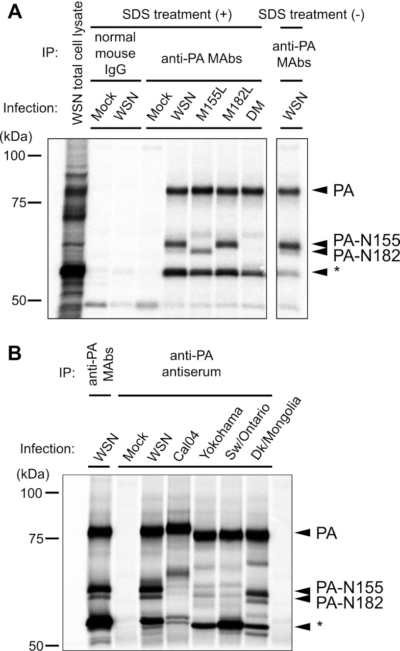
Fig 2.
Detection of N-truncated PAs in virus-infected cells. MDCK cells were infected with WSN and PA mutant viruses (A) or several different influenza A virus strains (B) and at 3 h p.i. radiolabeled for 3 h in [35S]methionine/cysteine-containing medium. After cell lysis, the supernatants were immunoprecipitated (IP) with a mixture of mouse anti-PA monoclonal antibodies (MAbs) or rabbit anti-PA antiserum and analyzed by SDS-PAGE, followed by visualization by autoradiography. The asterisk indicates a fourth band whose nature remains unknown.

