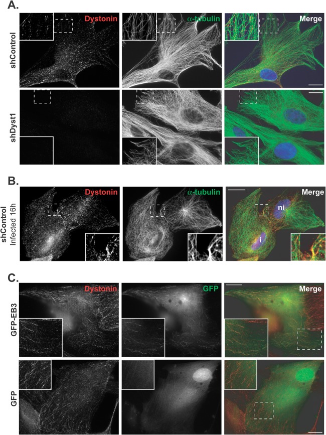
Fig 3.
Dystonin localization in HFFF2 cells. (A) shControl or shDyst1 cells were fixed, and the distributions of dystonin (red) and alpha-tubulin (green) were determined using antibodies DST and GAR568 and DM1A and GAM488, respectively. Identical exposure times were used to collect images of shControl and shDyst1 cells. (B) shControl cells were infected with vUL35RFP1D1, a virus that encodes a VP26 capsid protein fused to the mRFP. Sixteen hours postinfection, cells were fixed and stained as described above, except that secondary antibodies were GAR488 and GAM647. Infected cells (i) were distinguished from noninfected cells (ni) through mRFP fluorescence (not shown). (C) HFFF2 cells were transfected with pGFP-hEB3, which encodes GFP fused to the plus-end-binding protein EB3 or with pEGFP-C1 as a control. Twenty-four hours after transfection, cells were fixed and immunostained for dystonin as described for panel A (red). GFP-EB3 and GFP are visualized through autofluorescence (green). An area from each cell image (dashed box) is enlarged. Scale bars, 20 μm.

