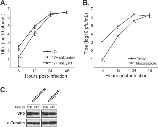
Fig 4.

Growth kinetics of HSV-1 in the absence of dystonin or of MTs. Single-step growth curves of HSV-1 WT virus (17+) in control cells or cells silenced for dystonin (A) or in the presence or absence of nocodazole (B). (A) HFFF2 cells were transduced with shControl or shDyst1 shRNAs or untreated before being infected with 5 PFU/cell of HSV-1 17+. One hour after infection, the viral suspension was removed and cells were acid washed. At 6, 12, 24, or 48 h after infection, cells were scraped into the medium and pelleted. The supernatant was harvested and the titer of progeny virus was determined on Vero cells. (B) HFFF2 cells were incubated with 10 μM nocodazole 1 h before infection and all the time of infection (nocodazole) or not at all (DMSO). Infection and titration were done as described for panel A. Experiments were done in triplicate. Error bars indicate standard deviations of the mean (SDM). Asterisks indicate statistical differences with control conditions (17+ in untreated cells in panel A and DMSO condition in panel B) as determined by a paired Student t test (*, P < 0.05; **, P < 0.01). (C) Western blot analysis of lysates obtained from shControl or shDyst1 cells infected with 5 PFU/cell of 17+ for the indicated times (16 h or 24 h pi). The progress of infection was visualized using the DM165 antibody directed against the major capsid protein VP5, and alpha-tubulin levels were monitored as loading controls using the DM1A antibody.
