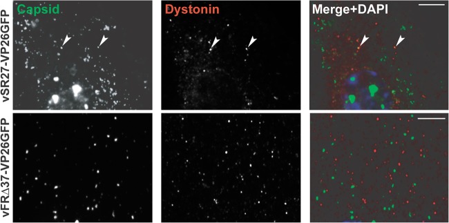
Fig 5.

Capsid association with dystonin. HFFF2 cells were infected with 1 PFU/cell of vSR27-VP26GFP or vFRΔ37-VP26GFP. Sixteen hours later, cells were fixed and endogenous dystonin was visualized using specific antibodies DST and GAR568 (red). Capsids were visualized through autofluorescence (green). Arrowheads indicate capsids colocalizing with dystonin spots. Scale bars, 5 μm.
