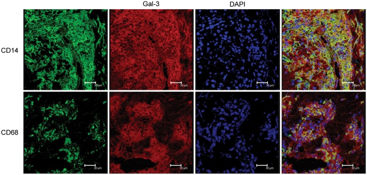
Figure 2.
Coexpression of macrophage markers with galectin-3 (Gal-3) expression in lepromatous leprosy (L-lep) skin lesions. Skin lesions from L-lep patients were sectioned and immunolabeled with monoclonal antibodies as indicated and visualized by confocal laser microscopy. Images were photographed using a ×63 objective. Scale bars = 30 μm. CD14 and CD68 were visualized as green fluorescence, and Gal-3 (9C4) was visualized as red fluorescence. Nuclei were labeled with DAPI. Coexpression (yellow) revealed the relatively close proximity of Gal-3 to macrophage markers CD14 and CD68.

