Figure 4.
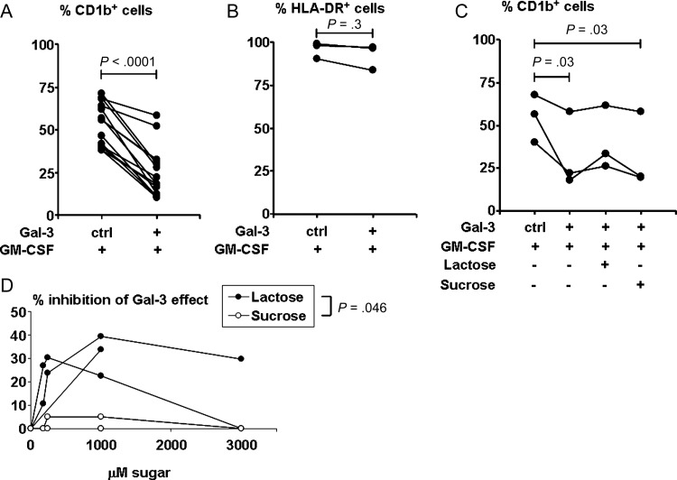
Effect of galectin-3 (Gal-3) on granulocyte macrophage colony-stimulating factor (GM-CSF)–treated monocytes. CD1b and HLA-DR expression on monocytes treated with medium or GM-CSF in the presence of Gal-3 or control buffer for 2 days as measured by flow cytometry is shown. Cell surface protein expression levels are expressed as the percentage positive for CD1b from 14 donors (A) and HLA-DR from 3 donors (B). Monocytes treated with medium alone expressed CD1b within a range of 0.3% to 14.5%, and HLA-DR within a range of 81.5% to 98.3%. Statistical analysis was performed using paired t test (A) and sign test (B), which is a nonparametric version of the paired t test. C, CD1b expression on monocytes treated as in (A) from 3 donors, with the addition of lactose or sucrose (1 mM each). Statistical analysis was performed with general linear model using Tukey method for multiple comparison adjustment. D, Dose titration of sugar inhibition of the effect of Gal-3 on CD1b expression. Results shown are from the same 3 donors as in (C). Values are reported as the percentage of decrease in the percentage of CD1b-positive cells induced by GM-CSF: [|(percent in the presence of Gal-3 minus percent in the presence of Gal-3 and sugar)|/|(percent in the presence of Gal-3 minus percent in the absence of Gal-3)|] ×100%. Statistical analysis was performed using mixed models assuming compound symmetry covariance matrix among repeated measures over several concentrations.

